Your thyroid ultrasound and TI-RADS, explained
Case of a fictitious patient: Héctor’s ultrasound report arrived on his patient portal on a Friday night: “1.7 cm solid, hypoechoic nodule, TR4.” His appointment was not until Tuesday. He read the report a dozen times. “TR4 sounded like stage 4,” he said later. It is not. Here is what those words mean.
Why ultrasound is the key test
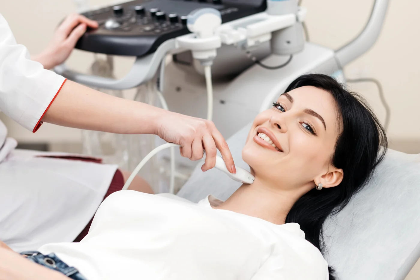
Ultrasound uses sound waves, with no radiation, to show the inside of the thyroid in detail. It shows the size of a nodule and, more importantly, its features, which help estimate the chance that it is cancer. Most nodules are benign, and ultrasound helps decide which few need a biopsy [1][4].

The words in your report
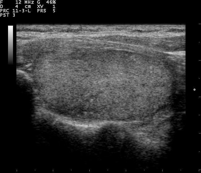
Radiologists describe nodules with a standard vocabulary [1]:
- Composition: cystic (fluid), spongiform (many tiny fluid spaces), mixed, or solid. Fluid-filled and spongiform nodules are reassuring.
- Echogenicity: how bright or dark the nodule looks compared with normal thyroid. “Hypoechoic” means darker. Very dark nodules are more concerning.
- Shape: “taller than wide” (when viewed in cross-section) is a concerning feature.
- Margins: smooth edges are reassuring; irregular edges, or growth outside the thyroid, are concerning.
- Echogenic foci: bright spots. Some (large “comet-tail” artifacts) are reassuring; tiny bright dots (“punctate echogenic foci”) are more concerning.
What the TI-RADS categories mean
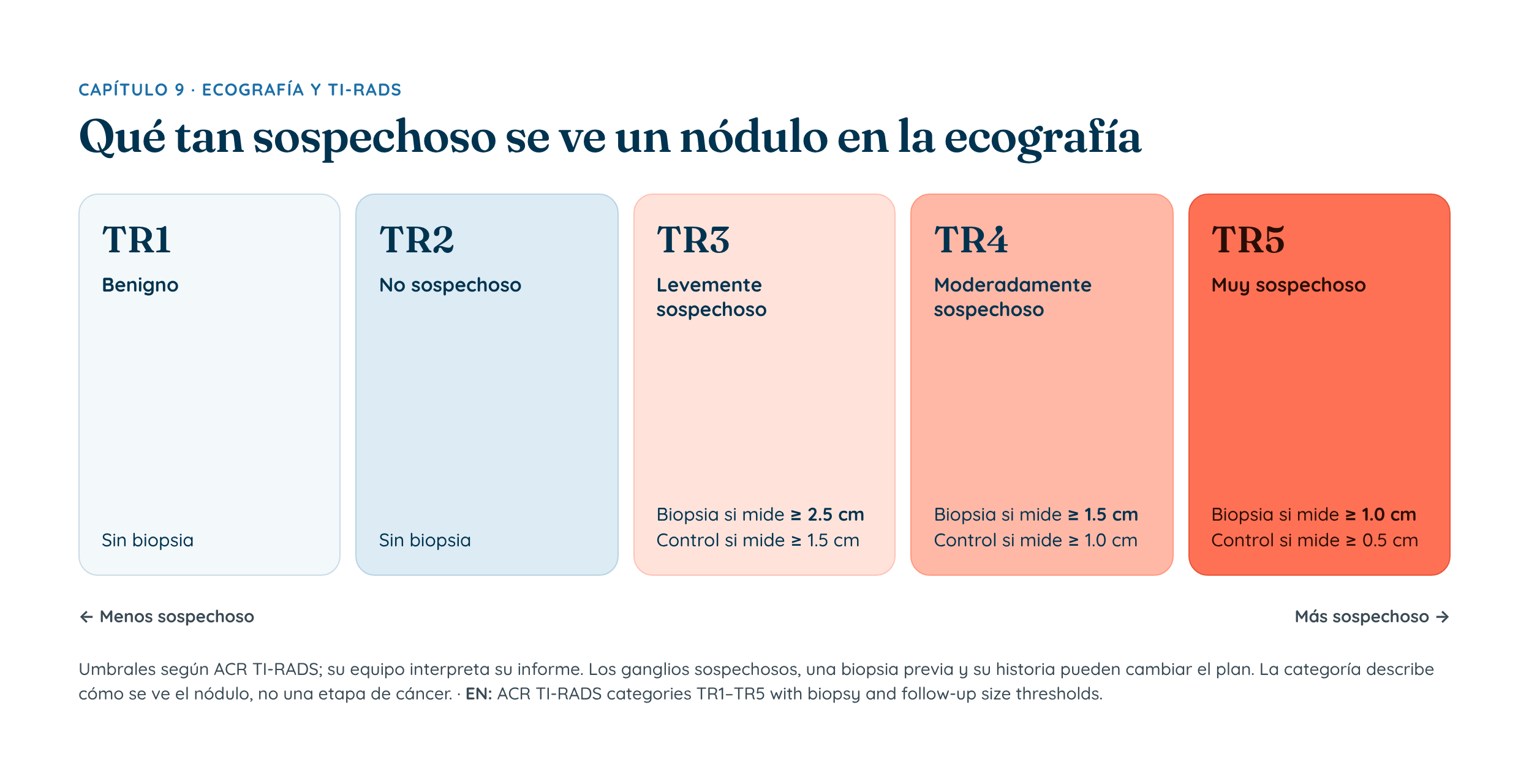
The American College of Radiology’s TI-RADS system adds up these features into a category from TR1 to TR5 [1]:
| Category | What it means |
|---|---|
| TR1 | Benign appearance |
| TR2 | Not suspicious |
| TR3 | Mildly suspicious |
| TR4 | Moderately suspicious |
| TR5 | Highly suspicious |
A TI-RADS category is not a cancer stage. TR4 means “moderately suspicious appearance,” not “stage 4.” Many TR4 nodules turn out to be benign; a biopsy is what tells.
Category plus size decides the next step
TI-RADS combines the category with the nodule’s size to recommend one of three things: no further action, ultrasound follow-up, or a fine-needle biopsy (Chapter 10) [1]. Small nodules, even with some concerning features, often only need follow-up. Larger nodules in higher categories are biopsied.
| Category | Biopsy usually considered at | Follow-up ultrasound at |
|---|---|---|
| TR1–TR2 | Not needed | Not needed |
| TR3 | 2.5 cm or larger | 1.5 cm or larger |
| TR4 | 1.5 cm or larger | 1.0 cm or larger |
| TR5 | 1.0 cm or larger | 0.5 cm or larger |
The size cut-offs above are the American College of Radiology (ACR) TI-RADS thresholds [1]. Suspicious-looking lymph nodes, a previous biopsy, your history and other factors can change what your doctor recommends.
This approach works: large reviews show ACR TI-RADS finds most cancers while avoiding many unnecessary biopsies [2][3].
What about artificial intelligence?
AI tools that analyze ultrasound images are being studied and show promise, especially in helping less experienced readers and reducing unnecessary biopsies [5]. For now they are a support for, not a replacement for, an experienced physician.
Questions to ask
- What is my nodule’s TI-RADS category and size?
- Based on that, do I need a biopsy, a follow-up ultrasound, or nothing more?
- When should the next ultrasound be?
- Were any lymph nodes in my neck abnormal?
Key points
- Ultrasound is the main test for thyroid nodules.
- TI-RADS categories (TR1–TR5) describe how suspicious a nodule looks, not a cancer stage.
- Category plus size decides whether you need a biopsy, follow-up or nothing.
- Discuss your report with your doctor rather than trying to score it yourself.
A head and neck surgeon experienced in thyroid and voice care can help.
Sources
- Tessler FN, et al. ACR Thyroid Imaging, Reporting and Data System (TI-RADS): White Paper of the ACR TI-RADS Committee. J Am Coll Radiol. 2017;14(5):587-595. doi:10.1016/j.jacr.2017.01.046 PubMed PMID 28372962
- Li W, et al. Diagnostic Performance of American College of Radiology TI-RADS: A Systematic Review and Meta-Analysis. AJR Am J Roentgenol. 2021;216(1):38-47. doi:10.2214/AJR.19.22691 PubMed PMID 32603229
- Castellana M, et al. Performance of Five Ultrasound Risk Stratification Systems in Selecting Thyroid Nodules for FNA. J Clin Endocrinol Metab. 2020;105(5):dgz170. doi:10.1210/clinem/dgz170 PubMed PMID 31690937
- Haugen BR, et al. 2015 American Thyroid Association Management Guidelines for Adult Patients with Thyroid Nodules and Differentiated Thyroid Cancer. Thyroid. 2016;26(1):1-133. doi:10.1089/thy.2015.0020 PubMed PMID 26462967
- De Cicco R, et al. Artificial intelligence-based systems as auxiliary tools for thyroid nodule malignancy detection on ultrasound: a systematic review and meta-analysis of diagnostic accuracy. Gland Surg. 2026;15(7):186. doi:10.21037/gs-2026-1-0126 PubMed PMID 42591757
This guide is for general education only. It does not diagnose conditions or replace a medical evaluation. Talk with your own doctor about your situation. In an emergency, call 911 now (in Puerto Rico or the United States; elsewhere, your local emergency number).